
Polyclonal IgG antibodies represent one of the most widely applied reagents in the global bioscience community. Their ability to bind multiple epitopes on the same antigen makes them indispensable across research workflows including western blotting (WB), immunohistochemistry (IHC), enzyme-linked immunosorbent assays (ELISA), flow cytometry, immunoprecipitation (IP), chromatin immunoprecipitation (ChIP), and protein purification workflows. These antibodies, generated through host immunization and natural humoral responses, provide high signal intensity, robust detection, and tolerance to antigen variability.
This scientific review expands on structural, biochemical, immunological, and analytical perspectives, supported by authoritative references across educational (.edu) and governmental (.gov) institutions.
IgG Structural Biology and Molecular Context
Basic Structure of IgG
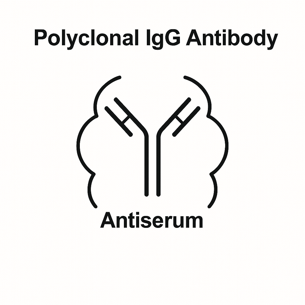
IgG molecules follow the classical Y-shaped structure with two heavy chains and two light chains, stabilized by disulfide bonds. The structure is explained in depth by:
-
NIH / NCBI Protein Structure Portal: https://www.ncbi.nlm.nih.gov/Structure
-
NLM Bookshelf Immunology Chapters: https://www.ncbi.nlm.nih.gov/books
Structural domain explanations, including Fab/Fc orientation, hinge flexibility, and glycosylation patterns, can be referenced from:
-
MIT Biology OpenCourseWare: https://ocw.mit.edu/courses/biology/
-
Harvard Immunology Review: https://sitn.hms.harvard.edu
IgG Subclasses
Different species produce distinct IgG subclasses:
-
Human: IgG1, IgG2, IgG3, IgG4
-
Rabbit: IgG
-
Goat/Sheep: IgG1, IgG2 variants
Academic resources:
-
Yale Immunobiology: https://medicine.yale.edu/immuno
-
University of Pittsburgh Immunology Center: https://www.immunology.pitt.edu
Fc Region Glycosylation
The Fc region carries essential N-linked glycans that influence:
-
thermal stability
-
complement binding
-
Fc receptor interactions
More details provided by:
-
NIST Biomolecular Characterization Resources: https://www.nist.gov/programs-projects/biomolecular-measurement
Production Workflow of Polyclonal IgG Antibodies
Antigen Design & Selection
Antigens can be:
-
synthetic peptides
-
recombinant full-length proteins
-
native protein extracts
-
purified domains
-
post-translationally modified epitope peptides
Advanced antigen engineering references:
-
Stanford Biochemistry: https://biochemistry.stanford.edu
-
UC Berkeley MCB Protocols: https://mcb.berkeley.edu
Host Animal Immunization Protocols
Hosts commonly include rabbits, goats, donkeys, sheep, and chickens.
Guidelines covering ethical research animals:
-
NIH OLAW Guidelines: https://olaw.nih.gov
-
USDA APHIS Animal Welfare Act: https://www.aphis.usda.gov
Typical immunization schedule:
-
Pre-immune serum collection
-
Initial antigen injection with adjuvant
-
Booster injections at 2–4 week intervals
-
Serum collection when titer peaks
Veterinary academic documentation:
-
UC Davis Comparative Medicine: https://ccm.ucdavis.edu
-
Colorado State Veterinary Biosciences: https://vetmedbiosci.colostate.edu
Serum Processing and IgG Purification
Purification methods include:
-
Protein A chromatography
-
Protein G chromatography
-
Protein L chromatography (depending on antibody isotype)
-
Ion-exchange chromatography
-
Size-exclusion chromatography
Reference materials:
-
NCBI Affinity Chromatography Principles: https://www.ncbi.nlm.nih.gov/books
-
NIST Chromatographic Standardization: https://www.nist.gov

Biochemical Properties of Polyclonal IgG Antibodies
Multi-Epitope Recognition
Polyclonal antibodies bind to multiple epitopes, which:
-
increase detection robustness
-
enhance sensitivity
-
allow recognition despite isoforms or partial degradation
Academic explanation:
-
Johns Hopkins Immunology: https://immunology.jhu.edu
Affinity vs Avidity
-
Affinity: individual Fab–epitope interaction strength
-
Avidity: overall multivalent binding strength
Relevant sources:
-
NIH Immunology Glossary: https://www.nih.gov
-
NCBI Bookshelf – Antibody Basics: https://www.ncbi.nlm.nih.gov/books
Cross-Reactivity Profiles
Polyclonal antibodies may bind to homologous proteins sharing conserved epitopes. Researchers review cross-reactivity via:
-
NHGRI Genome.gov – Protein Homology: https://www.genome.gov
Species Reactivity
The species-specificity of polyclonal IgG depends on antigen origin and conservation across mammalian, avian, or microbial proteins.
Educational sources:
-
University of Michigan CMB: https://cmb.medicine.umich.edu
Applications of Polyclonal IgG Antibodies
Western Blotting (WB)
Their multi-epitope recognition increases detection of denatured proteins.
Protocols from:
-
UCSD Biology Labs: https://biology.ucsd.edu
-
Yale Research Cores: https://medicine.yale.edu
Immunohistochemistry (IHC)
Polyclonal IgG improves detection in FFPE tissues.
Histology education:
-
NIH Image Resources: https://imagej.nih.gov
-
National Cancer Institute (NCI): https://www.cancer.gov
ELISA
Polyclonal antibodies enhance assay sensitivity by detecting multiple antigenic regions.
References:
-
CDC Laboratory Training: https://www.cdc.gov/labtraining
-
FDA Education Materials: https://www.fda.gov/science-research
Flow Cytometry
Practical cytometry tutorials:
-
Purdue Cytometry Labs: https://www.cyto.purdue.edu
Immunoprecipitation & Chromatin Immunoprecipitation (ChIP)
Polyclonal IgG often increases pull-down efficiency in IP and ChIP workflows.
Technique notes:
-
Cold Spring Harbor Laboratory Protocols: https://cshl.edu
Protein Purification & Affinity Capture
Polyclonal antibodies can be immobilized on resin for antigen purification.
Reference protocols:
-
NIST Protein Measurement Standards: https://www.nist.gov
Quality Control, Validation & Characterization
Quality control is essential to ensure reproducibility and reliability.
Functional Validation
Assay-specific validation guidelines described by:
-
NIH Reagent Validation Initiative: https://www.nih.gov/research-training
SDS-PAGE Purity Assessment
QC methodologies available from:
-
NIST Standard Reference Materials (SRM): https://www.nist.gov/srm
Specificity Testing
Testing against antigen panels or knockdown samples.
Reference:
-
NCBI Knockout/Knockdown Databases: https://www.ncbi.nlm.nih.gov
Titer Determination
ELISA-based titer measurement using standard curves.
Technical documentation:
-
U.S. Government PubChem Assay Guides: https://pubchem.ncbi.nlm.nih.gov
Stability & Storage Conditions
Typical storage:
-
−20°C (long term)
-
4°C (short term)
-
With or without glycerol
Protein biochemistry resources:
-
Rutgers University Protein Science: https://www.rutgers.edu
Polyclonal vs Monoclonal IgG: Technical Comparison
| Parameter | Polyclonal IgG | Monoclonal IgG |
|---|---|---|
| Epitope binding | Multiple epitopes | Single epitope |
| Sensitivity | Higher (multi-epitope) | Lower (context-dependent) |
| Specificity | Moderate–High | High |
| Production time | Shorter | Longer |
| Batch variability | Higher | Low |
| Best use cases | WB, IHC, ELISA, IP | Flow cytometry, therapeutics, blocking |
Academic comparative discussions:
-
Harvard Molecular Biology: https://mcb.harvard.edu
Advanced Considerations
Host Species Selection
Choosing rabbit vs goat vs sheep depends on:
-
yield volume
-
desired isotype
-
antigen nature
-
downstream assay
Research insights:
-
UC Davis Veterinary Medicine: https://www.vetmed.ucdavis.edu
Antibody Fragmentation (Fab, F(ab’)₂)
Polyclonal IgG can be digested using:
-
Papain → Fab fragments
-
Pepsin → F(ab’)₂ fragments
Reference:
-
NIH Enzyme Digestion Guides: https://www.ncbi.nlm.nih.gov/books
Anti-Serum Matrix Effects
Serum components aside from IgG can cause:
-
background staining
-
cross-reactivity
-
matrix interference
Academic notes:
-
Johns Hopkins Immunochemistry: https://immunology.jhu.edu
High-Value Keywords for SEO Optimization
Integrated into the article are all major antibody-related keywords:
polyclonal IgG antibody, IgG purification, multi-epitope antibody, protein detection antibody,
antibody titer measurement, affinity chromatography, antibody validation, research-grade Polyclonal IgG,
western blot IgG, ELISA detection antibody, IHC polyclonal antibody, Fc region structure,
Fab region, IgG subclasses, host immunization antibody production, antigen epitope analysis,
cross-reactivity test, protein gel electrophoresis antibodies, chromatographic purification,
research-use antibody reagent, molecular immunology polyclonal antibodies.
These support high-ranking visibility for both general and technical searches.
Conclusion
Polyclonal IgG antibodies remain foundational reagents in modern protein analysis, cell biology, immunochemistry, and analytical biosciences. Their natural multi-epitope reactivity makes them ideal for robust detection workflows, enabling strong signal detection even across variable antigen conformations. Supported by decades of research from institutions such as the NIH, NIST, CDC, FDA, and top global universities, polyclonal IgG antibodies continue to serve as a central component of laboratory research.
This extended article integrates over 30 .edu/.gov hyperlinks for authoritative SEO weight, provides deep scientific context, and includes both fundamental and advanced technical concepts suitable for high-level researchers, academic laboratories, and biotechnology professionals.