
Introduction
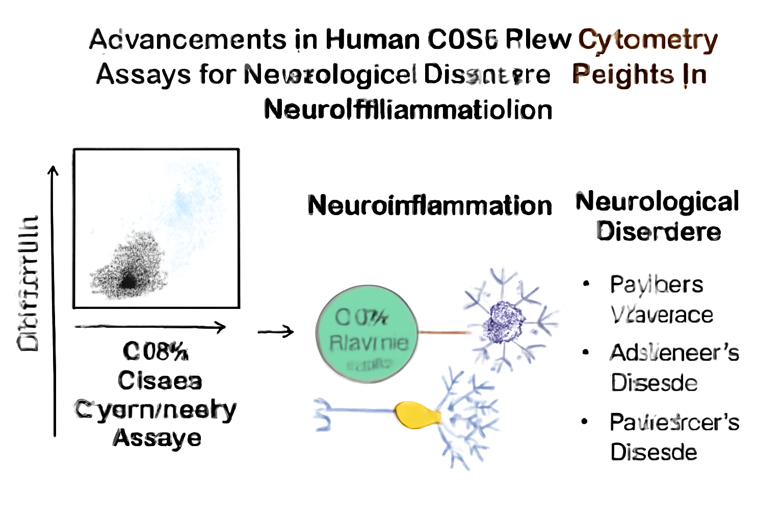
Neuroinflammation is a common pathological feature in many neurological disorders. The immune system’s involvement in the brain, often through microglia and immune cells, has led to the growing recognition of the role inflammation plays in neurodegeneration. CD56 (also known as neural cell adhesion molecule, NCAM) is a key marker in immune cells, particularly natural killer (NK) cells, which are crucial in neuroinflammatory responses. Flow cytometry, a powerful technique for analyzing cell populations, has made substantial progress in detecting and quantifying CD56-positive cells within the brain, offering new insights into immune involvement in diseases like multiple sclerosis (MS), Alzheimer’s disease (AD), and Parkinson’s disease (PD).
This article will explore the recent advancements in human CD56 flow cytometry assays, highlighting their importance in understanding neuroinflammation, particularly in the context of neurological disorders. It will also discuss how these innovations are shaping research and therapeutic strategies.
CD56 and Its Role in Neuroinflammation
CD56 is a cell adhesion molecule that plays a critical role in the immune response, especially in the brain. It is expressed on NK cells, a type of innate immune cell that is vital for maintaining immune homeostasis in the central nervous system (CNS). NK cells have been linked to neuroinflammation in various neurological conditions. In the context of diseases like multiple sclerosis, Alzheimer’s disease, and Parkinson’s disease, CD56-positive NK cells are often found in high concentrations at sites of inflammatory lesions.
-
Multiple Sclerosis (MS): CD56+ NK cells contribute to myelin damage by infiltrating into the CNS, where they mediate the destruction of oligodendrocytes. This results in the loss of the myelin sheath, causing impaired neural conduction and ultimately, the characteristic symptoms of MS, such as motor and cognitive dysfunction (National MS Society – Neuroinflammation in MS).
-
Alzheimer’s Disease (AD): In AD, the accumulation of amyloid plaques triggers chronic inflammation, which involves CD56+ NK cells. These cells are involved in the activation of microglia—the resident immune cells of the CNS—leading to synaptic dysfunction and neuronal death, hallmarks of the disease (Alzheimer’s Disease Neuroimaging Initiative – Inflammation in AD).
-
Parkinson’s Disease (PD): In Parkinson’s, CD56+ NK cells have been shown to interact with dopaminergic neurons, contributing to neuronal damage. The activation of neuroinflammation in the substantia nigra region of the brain has been linked to the degeneration of these neurons, resulting in motor deficits (Parkinson’s Foundation – Neuroinflammation in PD).
Innovations in CD56 Flow Cytometry Assays
Flow cytometry assays have traditionally been used to analyze cell populations based on surface markers such as CD56. Recent advancements in the technology have vastly improved the resolution and analytical power of these assays, enabling more precise and detailed investigation of CD56+ NK cells in neuroinflammatory diseases. These innovations are now making it possible to gain deeper insights into the mechanisms of immune cell activity in the brain.
-
High-Throughput and Multiparameter Flow Cytometry:
Modern flow cytometers now allow for multiparameter analysis, meaning that multiple surface and intracellular markers can be measured simultaneously. This technique enables the detection of subtle changes in immune cell phenotypes and activation states, even in rare populations such as CD56+ NK cells. By measuring multiple biomarkers, it is possible to correlate immune activation with disease progression, offering a more comprehensive view of neuroinflammatory dynamics. This approach has been particularly valuable in Alzheimer’s disease, where the inflammatory response is multifactorial and complex (PubMed – Multiparameter Flow Cytometry in Neuroinflammation). -
Advanced Cell Sorting Techniques:
The ability to isolate and sort CD56+ NK cells with high precision is crucial for studying the roles these cells play in neuroinflammation. Recent advancements in fluorescence-activated cell sorting (FACS) allow for more refined separation of immune subsets, enabling researchers to focus on specific populations of NK cells. This advancement has been instrumental in studying the interactions between CD56+ cells and microglia, key players in diseases like multiple sclerosis and Parkinson’s disease (Nature Immunology – Advances in FACS Technology). -
Integration with Single-Cell RNA Sequencing:
The integration of single-cell RNA sequencing with flow cytometry assays has enabled researchers to explore the transcriptomic profiles of CD56+ NK cells. By combining these techniques, it is now possible to examine gene expression at a single-cell level, providing insights into how CD56+ cells contribute to neuroinflammation at the molecular level. This integration has uncovered new therapeutic targets for diseases like Alzheimer’s and Parkinson’s, where gene expression profiles can be linked to specific immune responses (NIH – Single-cell RNA Seq Integration).
Applications of CD56 Flow Cytometry Assays in Neurological Diseases
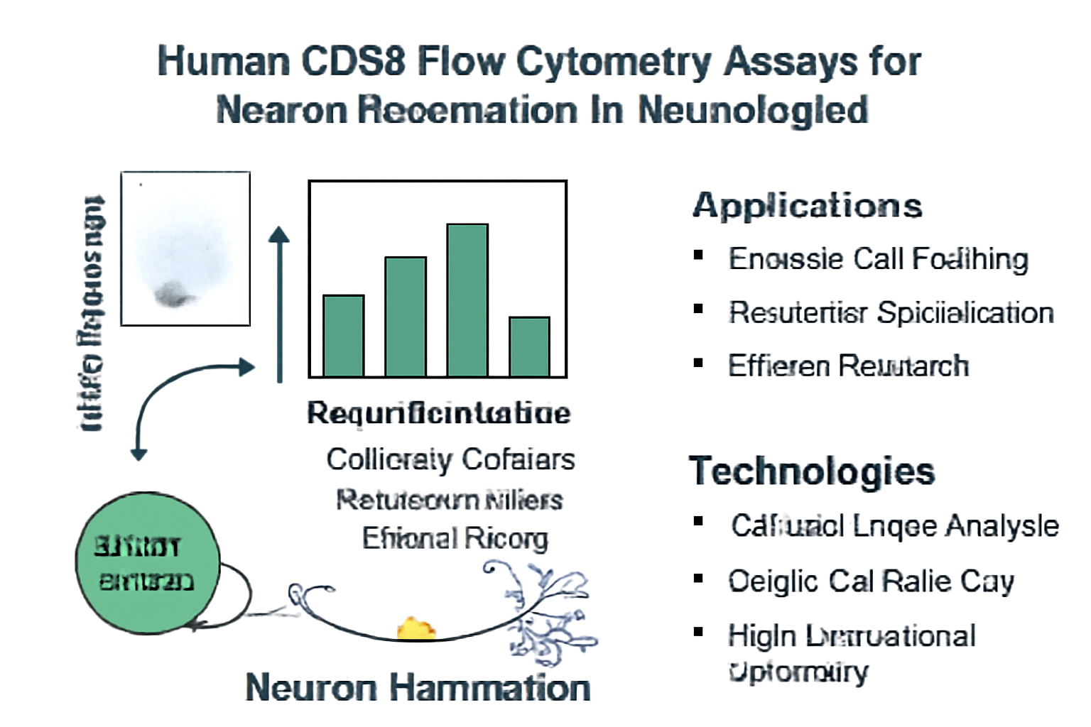
The application of CD56 flow cytometry assays in neurological research has greatly enhanced our understanding of neuroinflammation and its role in disease progression. Key areas of application include:
-
Monitoring Immune Infiltration in MS:
Flow cytometry allows for the precise quantification of CD56+ NK cells in cerebrospinal fluid (CSF) and peripheral blood of MS patients. By tracking changes in the number and activation state of these cells, researchers can monitor the progression of neuroinflammation and demyelination in real-time, providing valuable data for early diagnosis and prognosis (Multiple Sclerosis International Federation – Immune Cells in MS). -
Neuroinflammation in AD:
The use of CD56 flow cytometry assays has also provided valuable insights into the immune response in Alzheimer’s disease. By identifying and characterizing CD56+ NK cells in the brain and CSF, scientists can track the activation of these cells in response to amyloid beta deposition. This has led to a better understanding of the link between immune activation and cognitive decline (Alzheimer’s Association – Inflammation in Alzheimer’s). -
Parkinson’s Disease and Immune Response:
CD56+ NK cells have been implicated in the degeneration of dopaminergic neurons in Parkinson’s disease. Flow cytometry assays have helped to reveal the role of neuroinflammation in PD progression, especially how NK cells interact with other immune cells in the substantia nigra. These findings have paved the way for new research into immunomodulatory therapies for PD (Parkinson’s Foundation – Immunology in PD).
Technological Challenges and Future Directions
Despite significant advancements, there are still challenges that need to be addressed to fully leverage CD56 flow cytometry assays in neuroinflammatory research. Some of these challenges include:
-
Technical Complexity of Brain Tissue Analysis:
Brain tissue is highly complex, and isolating viable CD56+ NK cells from the CNS remains a technical challenge. The development of new techniques for isolating these cells from post-mortem brain tissue and CSF is crucial to advancing our understanding of neuroinflammation at the cellular level (Cell Reports – Challenges in Brain Tissue Cytometry). -
Data Analysis and Interpretation:
The large amount of data generated by high-dimensional flow cytometry poses a significant challenge in terms of data processing and interpretation. The integration of machine learning and artificial intelligence into data analysis workflows can help researchers uncover patterns in immune cell dynamics that were previously difficult to detect (Nature Methods – AI in Flow Cytometry). -
Improved Sensitivity and Specificity:
Increasing the sensitivity and specificity of CD56 detection in flow cytometry assays will be essential for identifying low-abundance populations of immune cells involved in neuroinflammation. Innovations in fluorescence techniques and nanotechnology may offer solutions to this problem (Nature Materials – Fluorescence in Cytometry).
Conclusion
The advent of human CD56 flow cytometry assays has revolutionized our ability to study neuroinflammation in neurological diseases. These advancements have allowed for more precise detection and characterization of immune cells, particularly CD56+ NK cells, in diseases like multiple sclerosis, Alzheimer’s, and Parkinson’s disease. As technology continues to evolve, the integration of single-cell RNA sequencing, multiparameter flow cytometry, and artificial intelligence will further enhance our ability to understand the immune mechanisms behind neurodegeneration and pave the way for new immunomodulatory therapies.
The future of neuroinflammatory research depends on continued innovation in flow cytometry technology, improved data analysis techniques, and collaborative efforts to identify novel biomarkers for early diagnosis and effective treatment strategies.