
Introduction
Fibrosis is a progressive biological process that involves the excessive accumulation of extracellular matrix (ECM) proteins, primarily collagen. In vitro fibrosis models serve as controlled platforms to study the cellular and molecular mechanisms underlying ECM deposition. These models are particularly valuable for screening antifibrotic agents, understanding tissue remodeling, and validating fibrotic signaling pathways.
Among the most essential analytical tools in these models are collagen assay kits. These kits allow researchers to measure collagen accumulation with precision, reliability, and scalability across various experimental conditions. The collagen content directly reflects the degree of fibrosis in cell cultures, organoids, or scaffold-based constructs.
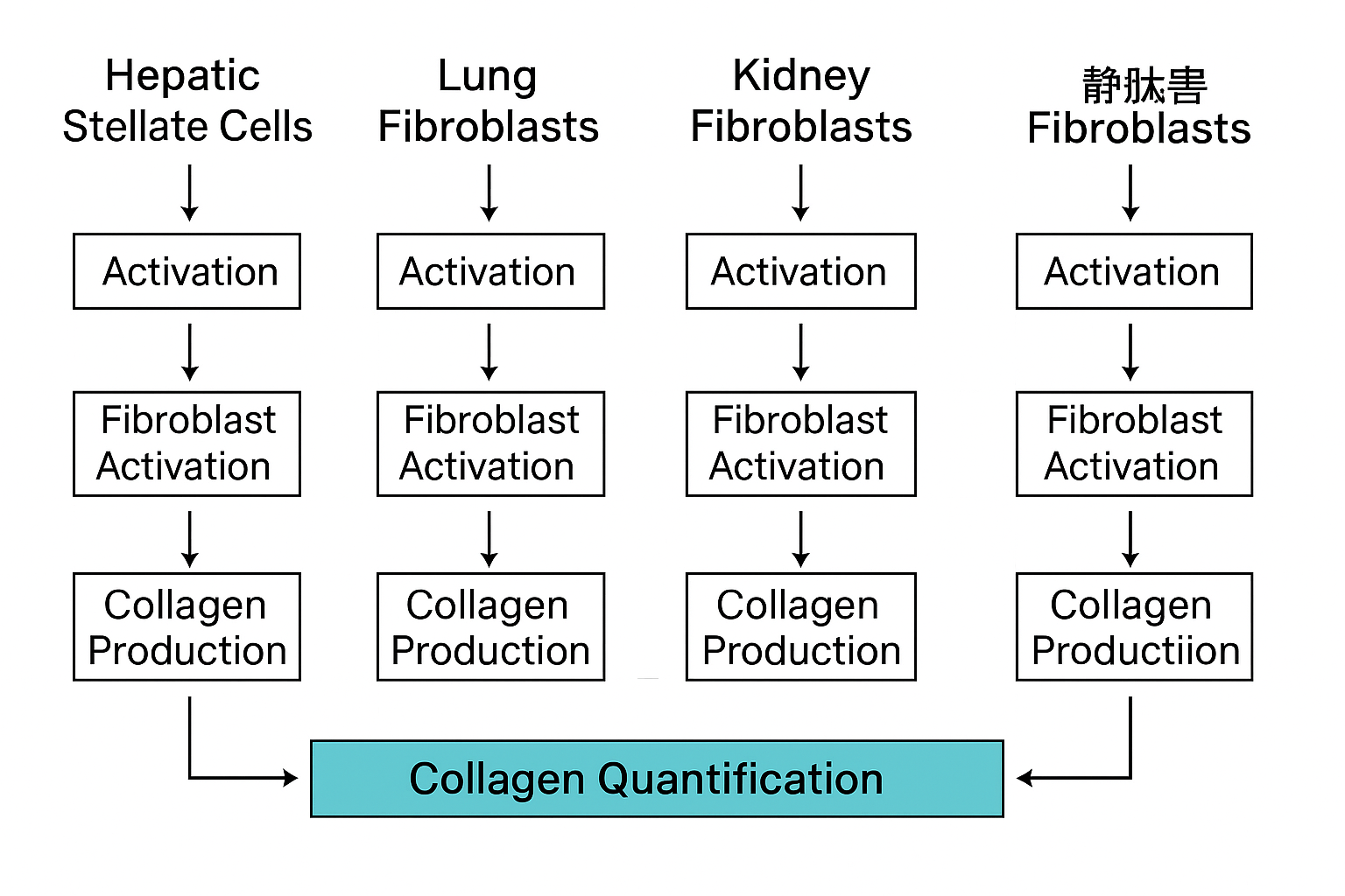
This article details the application of collagen assay kits in vitro fibrosis models using hepatic stellate cells, lung fibroblasts, kidney fibroblasts, cardiac fibroblasts, and dermal fibroblasts. It highlights quantification methods, technical considerations, and integration with other readouts, with references to verified educational and institutional resources.

Collagen and Its Central Role in Fibrosis
Collagen proteins are the primary structural components of the ECM. Among the 28 types identified, Type I and Type III collagen are most implicated in fibrotic diseases. In healthy tissues, collagen turnover is tightly regulated. Under pathological conditions, fibroblast-like cells become activated and secrete excessive collagen.
The shift toward a pro-fibrotic phenotype can be triggered by:
-
Cytokines (e.g., TGF-β1)
-
Mechanical stress
-
Oxidative stress
-
Chemical injury
Once activated, these cells remodel the ECM, leading to organ dysfunction. Monitoring collagen deposition in vitro is essential for dissecting these mechanisms.
For an overview of fibrosis-related research, refer to https://www.nhlbi.nih.gov and https://www.niddk.nih.gov.
Types of Collagen Assay Kits
Different formats of collagen assay kits are available, each suited to particular experimental goals:
a. Colorimetric Assays
-
Use dyes like Sirius Red that bind collagen.
-
Results are read spectrophotometrically.
-
Useful for total collagen in culture media or cell lysates.
b. Hydroxyproline Assays
-
Measure hydroxyproline, an amino acid abundant in collagen.
-
Often used after acid hydrolysis.
-
Reflects mature collagen content.
c. Fluorometric Assays
-
Utilize fluorescent probes that detect collagen directly.
-
Higher sensitivity compared to colorimetric methods.
d. ELISA-Based Assays
-
Use monoclonal antibodies to detect specific collagen isoforms.
-
Enable differentiation between COL1A1, COL3A1, and COL4A1.
Each type has its own advantages in terms of sensitivity, specificity, and ease of use. Standard protocols are available on https://www.ncbi.nlm.nih.gov/protocols.
Application in Specific In Vitro Fibrosis Models
a. Hepatic Stellate Cells (HSCs)
In liver fibrosis, quiescent hepatic stellate cells are activated into myofibroblasts that deposit type I collagen. This process is mimicked in vitro using:
-
LX-2 or primary human HSCs
-
Stimuli: TGF-β1, PDGF, ethanol metabolites
Collagen assay kits are applied to:
-
Quantify secreted collagen in culture media
-
Compare fibrotic potential of different compounds
-
Evaluate inhibition by antifibrotic agents
Liver fibrosis research protocols: https://www.niddk.nih.gov/research-funding

b. Lung Fibroblasts and Pulmonary Fibrosis
Lung fibroblasts are major ECM producers in pulmonary fibrosis. Models use:
-
MRC-5, IMR-90, or primary lung fibroblasts
-
Stimulation with TGF-β1 or silica
Collagen assays are critical to:
-
Quantify total collagen
-
Compare drug-treated vs untreated cells
-
Validate mRNA–protein expression correlation
For pulmonary models, refer to:
c. Renal Fibroblasts and Kidney Fibrosis
Kidney fibroblasts and proximal tubular epithelial cells are used to model interstitial fibrosis:
-
Triggered by Angiotensin II, hypoxia, or glucose
-
Co-culture models mimic tubulointerstitial interactions
Collagen assays support:
-
Detection of fibrotic shift
-
Time-course monitoring of ECM remodeling
-
Evaluation of antifibrotic agents (e.g., ACE inhibitors)
Additional kidney disease studies:
d. Cardiac Fibroblasts in Heart Remodeling
After myocardial infarction, cardiac fibroblasts become collagen-producing myofibroblasts. In vitro:
-
Cultures exposed to Ang II, TNF-α, or mechanical stretch
-
Measured collagen reflects ECM expansion and stiffness
Assay applications:
-
Quantifying fibrotic load
-
Comparing effects of β-blockers
-
Screening new signaling inhibitors
Cardiac research references:
e. Skin Fibroblasts and Dermal Fibrosis
Skin models of fibrosis use fibroblasts in 3D collagen gels to simulate dermal thickening:
-
Stimuli: TGF-β1, bleomycin, UV exposure
-
Endpoints: contraction, collagen content, α-SMA expression
Applications:
-
Study systemic sclerosis
-
Measure collagen contraction rate
-
Quantify ECM under anti-scarring treatments
NIH funding and protocols:
Technical Considerations for Assay Optimization
To ensure reproducibility and accuracy:
-
Standard curves using purified collagen (e.g., bovine)
-
Biological replicates for statistical significance
-
Normalization to cell number or total protein
-
Serum-free medium to reduce interference
Quality control sources:
Multiplexing with Molecular Readouts
Combining collagen assays with:
-
qRT-PCR for COL1A1, ACTA2, FN1
-
Western blot for total and phosphorylated proteins
-
Immunofluorescence to localize collagen deposition
-
Confocal imaging for spatial ECM analysis
Protocol references:
Scaling for High-Throughput Screening
In vitro fibrosis models are scalable to 96- or 384-well formats, enabling screening of libraries for fibrosis-modulating agents. Collagen assays are adaptable for robotic platforms.
-
Plate-based collagen detection
-
Automated imaging + assay integration
-
Software for multi-parametric scoring
HTS infrastructure:
Conclusion
Collagen assay kits provide a foundational method for studying fibrosis in vitro. Their versatility, precision, and compatibility with multiple model systems make them indispensable for:
-
Dissecting ECM biology
-
Validating disease models
-
Screening antifibrotic interventions
From hepatic stellate cells to lung fibroblasts, collagen assays support translational workflows and discovery pipelines. Combined with gene, protein, and imaging readouts, these kits help generate a comprehensive profile of fibrotic activity.